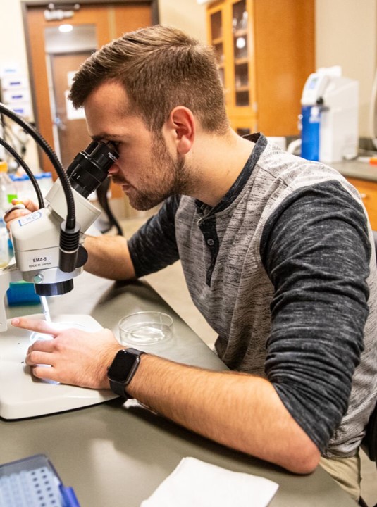
Brendin Flinn
B.S., Biological Sciences, Marshall University, 2022
Foci: EPR spectroscopy, microscopy, biochemistry, and molecular biology
Degree Sought: M.S. in Chemistry
Role:
Graduate Student Research Assistant
Date:
Summer 2022-Present
Funding:
National Science Foundation, Collaboration Grant
Extracurricular Interests: Competitive gaming, NCAA football, philosophy, music, theater, debate.
EPR Spectroscopy
One of Brendin’s principal interests is measuring the production of superoxide by tardigrades. To achieve this, electron paramagnetic resonance (EPR) spectroscopy is utilized. The technique is similar to nuclear magnetic resonance spectroscopy, but utilizes a continuous stream of microwave radiation with a sweeping magnetic field strength to detect paramagnetic species rather than nuclear spins (though nuclear spins are still observable in EPR spectra).
While superoxide is itself paramagnetic, its biological half-life is very small (on the order of milliseconds). In order to measure its production over time, Brendin employs an EPR spin probe called CMH. CMH reacts selectively and rapidly with superoxide to produce a nitroxide radical that is much more stable and still detectable via EPR, allowing for the indirect quantitation of superoxide production.


Slide 1 – EPR at Marshall University; Slide 2 – Reaction of CMH with Superoxide; Slide 3 – EPR Spectrum of the CMH Radical Used for Quantitation.
Microscopy
Brendin currently has two ongoing goals involving microscopy techniques: 1) Using confocal scanning laser microscopy (CSLM) to measure the volume of tardigrades in their various physiological states, and 2) Using scanning electron microscopy (SEM) to visualize morphological differences between cryptobiotes.
Volume Measurements
The basic principle behind Brendin’s work to measure the volume of tardigrades using CSLM is the utilization of a technique called shadow imaging. Brendin acquires many images at different focal planes (heights) to get a z-stack (set of images) that encompasses the whole tardigrade, but the image is a “shadow image” because the tardigrade appears dark while its surroundings appear bright. The z-stack can be analyzed in ImageJ, an image analysis software provided by the National Institutes of Health, to produce 3D renderings of the tardigrades. Those 3D renderings allow for accurate measurements of the volumes of the tardigrades.


3D Renderings of Tardigrades Produced from CSLM Z-Stacks. Slide 1 – Hydrated (non-stressed) tardigrade; Slide 2 – Sucrose-induced tun; Slide 3 – Sucrose-induced tun; Slide 4 – Hydrated tardigrade. All images acquired on the Leica SP5 at Marshall University (access provided through the Marshall University Molecular and Biological Imaging Center, MBIC).
Visualizing Morphological Differences
To visualize morphological differences between cryptobiotes, Brendin chose to use scanning electron microscopy to image the tardigrades. This is because normal optical techniques (like a normal benchtop microscope) make it difficult to see the fine morphological details of tardigrades due to the animals being mostly transparent. Scanning electron microscopy utilizes electrons instead of visible light to generate images and so allows us to visualize the small details of tardigrades (and also has the added benefit of being able to resolve much finer structures than light microscopy).


Scanning Electron Microscope Images of Tardigrades. Slides 1/2 – Sucrose-induced tuns. Slides 3/4 – CaCl2-induced tuns. All images acquired on the JEOL JSM 7200FLV SEM at Marshall University (access provided through the Marshall University Molecular and Biological Imaging Center, MBIC).
